Home »
Categories
Blog Articles and News
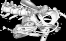
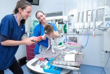
‘World first’ duplication case seen at Eastcott
Soft Tissue |
Our clinicians have been involved in a world first after successfully treating the first reported case of a dog with complete, unilateral urinary tract duplication. Tim Charlesworth, Head of Surgery at the multidisciplinary Eastcott Veterinary Referrals, in Wiltshire, has shared details of the remarkable case, which is believed to be unique. The dog in question…
Eastcott officially offering cats the gold-star treatment
Eastcott Blogs |
We are delighted to have been recognised as a Gold level cat clinic by the International Society of Feline Medicine (ISFM). The award is a big honour for the practice, with Eastcott Veterinary Referrals being recognised as delivering the highest standard of care and facilities for cats, further enhancing our reputation for first-class care and treatment. Eastcott…
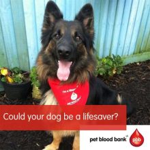
Pet Blood Bank donation session | Sunday 10th January
Eastcott Blogs |
Once again, we’re teaming up with Pet Blood Bank to provide a centre for them to perform regular blood donation sessions. Join us for our next session with Pet Blood Bank on Sunday 10th January. You do not need to be a registered client of Eastcott Vets for your dog to be able to help other patients. Your…
Christmas Opening Hours
Eastcott Blogs |
Please find below details of our opening times for the Christmas period Thursday 24th December Open as normal Friday 25th December Emergency and urgent referrals only. Please call 01793 528341 Saturday 26th December Sunday 27th December Monday 28th December Tuesday 29th December Open as normal Wednesday 30th December Thursday 31st December Friday 1st January Emergency…
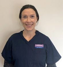
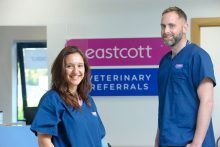
Top soft tissue specialist joins the team
Eastcott Blogs |
We are delighted to announce that highly qualified and experienced specialists soft tissue surgeon Faye Swinbourne has joined the team. Faye is an ECVS specialist in small animal surgery, RCVS specialist in small animal surgery (Soft Tissue) and EBVS® European specialist in small animal surgery. She will team up with fellow specialists Tim Charlesworth and Andrew…
Eastcott completes £3 million expansion
General Blog and News |
We’ve just completed an 18-month, £3 million extension and refurbishment programme at our practice on Dorcan Way which means we can now deliver healthcare and facilities to rival those enjoyed by humans.
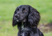
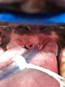
Maxillofacial surgery in 10-month-old Cocker Spaniel
Oral and Maxillofacial Surgery |
Lenny, a 10-month-old Working Cocker Spaniel, was sadly a little too inquisitive for his own good. Whilst exploring on a walk one day this young puppy startled a horse and received short shrift, in the form of a glancing blow to the jaw.
Launch of state-of-the-art neurology service
Eastcott Blogs, Neurology and Neurosurgery |
We’re delighted to now deliver a comprehensive Neurology service to treat patients with conditions of the brain, spinal cord and neuromuscular system.
Pet Blood Bank donation session – Wednesday 7th October
General Blog and News |
We’re teaming up with Pet Blood Bank to provide a centre for them to perform regular blood donation sessions.
BOAS surgery what’s involved?
Soft Tissue |
Soft Tissue Veterinary Surgeon Hannah Prestwood explains why certain dog breeds have breathing difficulties and what can be done to help them.